Specialty Contacts


What Is Keratoconus? Understanding This Complex Eye Condition
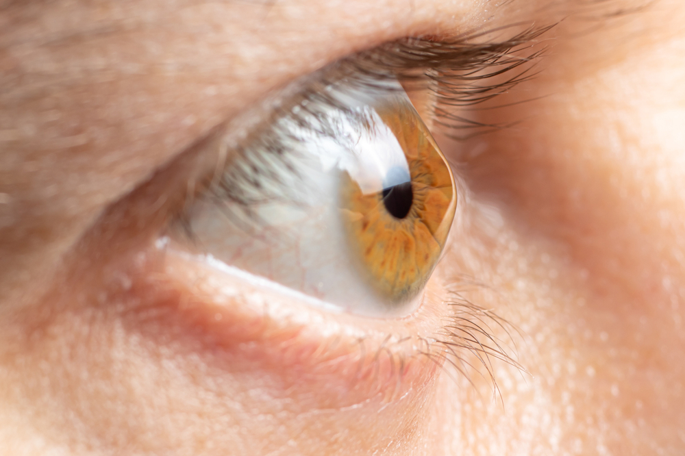
Keratoconus is an eye condition that causes the cornea to thin and gradually bulge into a cone-like shape. This irregularity distorts the way light enters the eye, leading to blurry or distorted vision that worsens over time. While the exact cause of keratoconus is not fully understood, genetic and environmental factors are believed to play a role.
Because keratoconus can significantly impact vision and daily activities, early diagnosis and specialized treatment, such as
custom contact lenses, are essential in managing the condition effectively. Understanding the symptoms, progression, and available treatment options can help individuals maintain clear, comfortable vision.
What Causes Keratoconus?
The exact cause of keratoconus is not fully understood, but several factors may contribute to its development, including:
• Genetics: A family history of keratoconus increases the risk of developing the condition.
• Frequent Eye Rubbing: Excessive rubbing of the eyes may contribute to corneal thinning and progression of keratoconus.
• Underlying Conditions: Some connective tissue disorders and inflammatory conditions have been linked to an increased risk of keratoconus.
The condition typically begins in the teenage years or early adulthood and progresses over time, sometimes stabilizing in later years.
Signs and Symptoms of Keratoconus
Keratoconus symptoms can vary depending on the severity of the condition. Common signs include:
• Blurred or distorted vision
• Increased sensitivity to light and glare
• Frequent changes in eyeglass prescriptions
• Difficulty seeing clearly at night
• Sudden worsening or clouding of vision in advanced cases
How Is Keratoconus Diagnosed?
Diagnosing keratoconus requires a comprehensive eye exam with specialized testing to evaluate the shape and thickness of the cornea. An optometrist may use corneal topography, a non-invasive imaging technique that maps the surface curvature of the cornea, revealing any irregularities or signs of steepening. Pachymetry is another diagnostic tool used to measure corneal thickness, which can help detect early thinning associated with keratoconus. Additionally, a slit-lamp examination allows the doctor to assess corneal health and identify signs such as scarring or stress lines (Vogt’s striae). Since keratoconus often progresses gradually, regular monitoring is crucial to track changes and determine the most effective treatment approach.
Specialty Contact Lenses for Keratoconus
As keratoconus progresses, standard eyeglasses and soft contact lenses may no longer provide clear vision. Fortunately, several specialty contact lens options are designed specifically to address the irregular corneal shape associated with keratoconus.
RGP lenses provide clear vision by creating a smooth optical surface over the irregular cornea. These lenses are smaller than soft contact lenses and offer better visual clarity in mild to moderate keratoconus.
Scleral lenses are larger-diameter gas permeable lenses that vault over the cornea and rest on the sclera (the white part of the eye). These lenses create a tear-filled space between the lens and the cornea, providing superior comfort and improving vision in moderate to severe keratoconus cases.
Some patients with mild keratoconus may benefit from custom soft lenses designed specifically to fit their irregular corneas. These lenses provide improved comfort while still offering some vision correction.
Managing Keratoconus for Long-Term Eye Health
Early detection and proper management are crucial in slowing the progression of keratoconus. In addition to specialty contact lenses, treatment options such as corneal cross-linking (CXL) can help strengthen the cornea and prevent further deterioration. In advanced cases, corneal transplants may be necessary to restore vision.
Schedule a Consultation at San Marcos Vision Center Today
At San Marcos Vision Center, we offer advanced diagnostic tools and specialized contact lens fittings to help manage keratoconus effectively.
If you or a loved one is experiencing symptoms of keratoconus, schedule a eye exam to explore the best vision solutions tailored to your needs. Visit our office in San Marcos, Texas, or call (512) 890-0660 to book an appointment today.


MAP
© 2026 San Marcos Vision Center. All Rights Reserved. Accessibility Statement - Privacy Policy - Sitemap
Powered by: